December Case of the Month: Navicular Cyst
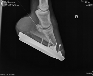
Molly became lame in her right front foot. She has had inconsistent lameness in that hoof previously that was managed with diligent care of her hoof angles, toe length and heel height. This time however, despite the appropriate farriery, she became and remained lame. Nerve blocks, numbing the sensation to her heel, confirmed the general location of her pain: her right front heel region.
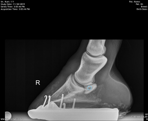
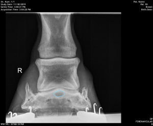
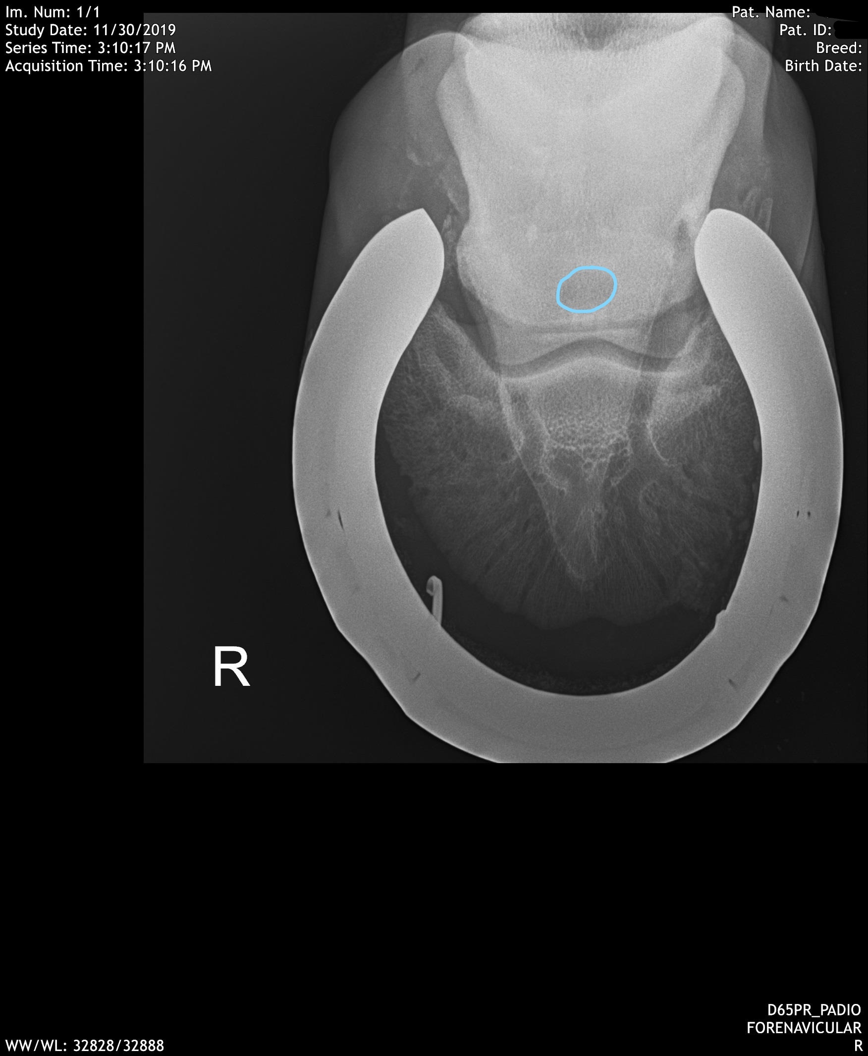
There can be multiple structures within the heel of the hoof that can be injured or diseased causing heel pain. These include, the deep digital flexor tendon, impar ligament, cruciate ligaments, navicular bursa, coffin joint, navicular bone, etc. Radiographs confirmed the abnormality was in Molly’s navicular bone, but, interestingly, it was not due to the common erosive or sclerotic (hardening) changes we see with Podotrochlear Syndrome (Navicular Syndrome). On radiographs, we can see a large cyst-like lesion within the navicular bone.
Bone cysts are fairly common findings in joints including the stifle, tarsal (hock), carpal (knee), fetlock, pastern and coffin joints, specifically involving OCDs. Many of these cysts develop when the horse is very young and is associated with abnormal cartilage development, and we term these cysts ‘subchondral bone cysts’. Molly’s cyst is more unique in that it is within the navicular bone. Molly was treated with coffin joint and navicular bursa injections. We recently received an update on Molly and she is very comfortable at the walk and trot and her owner is looking forward to Molly’s favourite activity, trail riding, when the weather gets better!